AngioAnalytics™ allows you to measure foveal avascular zone (FAZ) parameters, areas of abnormal flow in the outer retina and choroid, and vessel density of the superficial vascular complex, deep vascular complex, and radial peripapillary capillaries. Multi scan analysis enables assessment of visit-to-visit change, and trend reports predict change over time to bring powerful new information to your daily clinical practice. Watch the video.

AngioVueHD produces high-density scans with 73% more sampling points* to give you a 6x6mm scan with outstanding resolution. Now you can confidently evaluate fine vessels extending beyond the central 3x3mm region of the macula. Automatic montage (shown at right) expands the field of view even further by instantly combining macula and optic disc images for a 10x6mm widefield view.
*73% more sampling points than current AngioVue 6mm x 6mm scan

AngioVue® lets you see the retinal vessels in a whole new way. Three-dimensional images are separated into individual layers of retinal vasculature to show you an unprecedented level of detail and isolate areas of interest.1
1. Spaide RF, Klancnik Jr JM, Cooney, MJ. Retinal Vascular Layers Imaged by FA and Optical Coherence Tomography Angiography. JAMA Ophthalmol. 2015;133(1):45-50. Published online October 09, 2014.

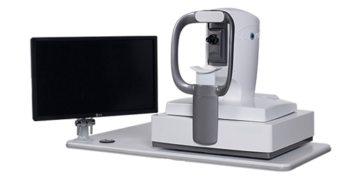
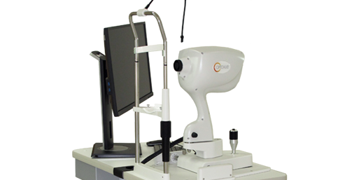
The AngioVue system is available in three configurations to meet the specific needs of your practice. AngioVue Comprehensive offers OCTA with extensive analytical functionality, while AngioVue Essential™ simplifies OCTA image interpretation with a single-page report. AngioVue Comprehensive and AngioVue Essential configurations include structural OCT for retina, glaucoma and anterior segment applications. AngioVue Retina combines OCTA with retina-only structural OCT. AngioVue Essential and Retina may be upgraded to AngioVue Comprehensive at any time.

The AngioVue Imaging System has been installed in more practices worldwide and featured in more peer-reviewed publications than any other OCTA technology. With Optovue, you can trust you’re partnering with a company committed to industry-leading clinical education, technical support and product innovation.

The AngioVue Imaging System uses the proprietary SSADA algorithm to produce stunningly detailed images and to minimize scan acquisition time. Optovue’s exclusive Motion Correction Technology (MCT™) reduces motion artifact, while 3D Projection Artifact Technology greatly improves OCTA image quality and enables accurate measurement and interpretation of OCTA images.
| Technical Specifications | |
| OCT scanning speed | 70,000 A-scans per second |
| Optical axial resolution | ~5 microns (digital pixel sampling = 3 μm) |
| Optical transverse resolution | ~15 microns |
| OCT axial imaging depth | 2 to 3 mm (dependent on scan protocol) |
| AngioVue imaging volume 3×3 mm | 304 x 304 A-lines (2 repeats / B-scan) |
| AngioVueHD imaging volume 6×6 mm, 4.5×4.5 mm | 400 x 400 A-lines (2 repeats / B-scan) |
| Acquisition time per imaging volume | (209K / 70K) ~3 seconds |
| AngioVue imaging size (retina) | 3×3, 6×6, 8×8 mm |
| AngioVue imaging size (optic disc) | 3×3, 4.5 x 4.5 mm |
| AngioVue imaging size (montage) | 10×6 mm |
| Recommended Networking Specifications | |
| Operating System | Windows 7, 8 and 10; 64-bit OS compatible |
| Processor Speed | 3.0 GHz; Intel Quad core (desktop); Core 2 (laptop) |
| Network Bandwidth | 1 Gbps or higher |
| Computer RAM | Minimum 8GB RAM |
| Recommended 16GB RAM | |
| Monitor Resolution | 1920 x 1080 at 32-bit color |
| Table Specifications | mm (in) |
| Width (W): | 950 (37.4) |
| Length (L): | 700 (27.6) |
| Height (H): | 690~890 (27.2~35.2) |
| Weight: | 65 KG (143 LB) |